Honouring Excellence
in dentistry

Odeon Theatre

Majestic Hall

Dresscode

Blacktie

December 6th, 2025

6:00 PM

PARTNERS

ACADEMIC

PLATINUM

INSTITUTIONAL

GOLd

Silver

rda logo negru

A prestigious gala where the most

accomplished and experienced dentists

from Romania and the Republic of Moldova are awarded.

Over the course of the three editions, we have recorded a total of:

ATTENDEES
0 +
JUDGED CASES
0 +
TROPHIES
0
PRIZES
0 K€+

Those prizes are awarded alongside the gratitude and respect we owe to the dentists who have chosen to present their most complex and interesting cases at the gala.

Odeon Theatre

Majestic Hall

dresscode

blacktie

December 6th, 2025

6:00 PM

Event Opening

Arrival and reception of participants

18:00 - 18:30

"camerata regală" concert

More details soon 

18:30 - 19:15

gala

The doctors who have reached the grand final will take the stage to present their cases, and the audience will vote for the winners

19:15 - 22:30

Ectopic reception

Networking and socializing, amuse-bouches and preosecco in a distiguisehd atmosphere in the theatre foyer

22:30 - 00:00

*The registration fee for a case includes ONLY ONE invitation to the event.

Cu sprijinul

Premii oferite de:

Sunt acceptate la această categorie doar restaurările directe din compozit ce respectă cerințele din descriere. Sunt acceptate atât restaurări directe pentru grupul dentar anterior, cât și pentru cel posterior. 


Descrierea cazului: 

  • Concurenții trebuie să includă o scurtă descriere a cazului clinic, care să cuprindă nu mai mult de 3500 de cuvinte 
 
  • Descrierile trebuie să conțină diagnosticul inițial, diagnosticul final, planul de tratament, materialele utilizate în conformitate cu protocolul de tratament și un scurt rezumat al cazului 
 
  • Prezentarea trebuie să cuprindă fotografii de tip before/after ± documentație radiologică atât de inițiere a tratamentului, cât și de după încheierea acestuia 
 
  • În cazul în care au fost restaurate margini incizale (atunci când au fost restaurați și caninii), prezentarea trebuie să conțină fotografii cu mandibula în protruzie și laterotruzie 


Instrucțiuni privind documentarea foto a cazului înscris: 

  • Îndepărtați saliva, placa bacteriană sau orice rest de ciment restant 
 
  • Centrați camera într-un unghi adecvat 
 
  • Încercați să folosiți același fundal atunci când este necesar, se recomandă contrastoare 
 
  • Oglinzile, retractoarele și alte astfel de elemente nu ar trebui să se afle în câmpul vizual 
 
  • Imaginile trebuie să aibă claritate, contrast, luminozitate, calitate și focus bun pentru a se putea evidenția detaliile importante care pot aduce departajarea 
 
  • Fotografiile trebuie să fie simetrice și decupate corespunzător 

Only endodontic cases that meet the specified criteria are accepted. Diagnosis must align with the international nomenclature published by the American Association of Endodontists that can be found here


Case Description and Photo Documentation: 

  • Pre- and post-operative radiographs are mandatory and represent the minimum requirement for submission. Intra-operative images such as working length and cone-fit radiographs are recommended
 
  • All intra-operative X-rays and photos must show the use of rubber dam isolation (e.g., presence of clamp on X-ray; for multiple tooth isolation, an intraoral photo is required if clamps are not visible on the radiograph)
 
  • OPG/panoramic X-rays are not accepted. Required radiographs must be periapical, follow the parallel technique, and show the treated tooth centered along with the apical area and any pathology 
 
  • CBCT may be accepted if its use is justified 
 
  • Follow-up radiographs should be taken using the same incidence as the initial ones. For cases with apical pathology, follow-ups at 12 and 24 months that show healing will earn a higher score 
 
  • Clinical images may be taken with a microscope camera, macro/DSLR/APSC camera, smartphone, or any device that can provide detailed, focused images 
 
  • Images must be clear, properly lit, high-resolution, and focused 
 
  • Rubber dam use is mandatory; no sign of leakage is allowed. Use of liquid dam, Teflon, or other additional methods is acceptable. A minimum of three images is required: isolation, pre-obturation, and post-obturation 
 
  • If the clinician also performed core build-up before or after endodontic treatment (with or without post placement), this will also be evaluated 

Only indirect aesthetic restorative treatments that meet the specified criteria are accepted. Cases must involve restorations within the patient’s aesthetic zone. 


Case Description: 

  • A brief clinical case description, not exceeding 3,500 words 

  • Include initial and final diagnosis, treatment plan, materials used, and a concise summary 

  • Presentation must include before/after photos ± radiographic documentation from start to finish 

  • If incisal edges were restored (e.g., involving canines), photos of mandibular protrusion and laterotrusion must be included 


Photo Documentation Guidelines: 

  • Remove saliva, plaque, or cement residues 

  • Align the camera properly 

  • Use consistent background; contrastors recommended 

  • Avoid mirrors, retractors, or other tools in the frame 

  • Ensure high clarity, contrast, brightness, and focus 

  • Photos must be symmetrical and properly cropped 

Only multidisciplinary treatments that meet the specified criteria are accepted. Eligible cases must involve at least two dental specialties and likely a team that worked together on diagnosis, planning, and execution. A team is not mandatory if the clinician stays within the scope of practice defined by law and involves at least two specialties. 


Cases that only include implant insertion followed by prosthetic rehabilitation must be entered under Implant-Prosthetic Treatment. 


Case Description: 

  • Brief clinical case description, max 3,500 words 

  • Include initial and final diagnosis, treatment plan, materials used, and summary 

  • Must include before/after photos ± relevant radiographs 

  • If incisal edges were restored, photos in protrusion/laterotrusion are required 


Photo Documentation Guidelines: 

  • Remove all residues (saliva, plaque, cement) 

  • Center the camera appropriately 

  • Use the same background when applicable; contrastors recommended 
  • No retractors, mirrors in the frame 

  • High clarity, quality, and focus required 

  • Images must be symmetrical and correctly cropped 

Only cases that include both implant surgery and prosthetic stages are accepted. If other specialties are involved, the case must be submitted under Interdisciplinary Oral Rehabilitation. 


Case Description: 

  • A brief clinical case description, not exceeding 3,500 words 
 
  • Include initial and final diagnosis, treatment plan, materials used, and a concise summary 
 
  • Presentation must include before/after photos ± radiographic documentation from the start and completion of treatment 
 
  • If incisal edges were restored (e.g., involving canines), images in mandibular protrusion and laterotrusion must be included 


Photo Documentation Guidelines: 

  • Remove all visible saliva, plaque, or residual cement 
 
  • Properly align the camera 
 
  • Use a consistent background when necessary; contrastors are recommended 
 
  • Avoid including mirrors, retractors, or other elements in the visual field


  • Photos must be clear, well-lit, in focus, and of high quality 


  • Images must be symmetrical and appropriately cropped 

Only treatments carried out entirely under a digital workflow are accepted, in accordance with the category’s criteria. This category is open to all clinical cases that follow a complete digital protocol. The main requirement is that every step of the treatment be executed using digital technologies (scanner, CAD/CAM, etc.). 


Case Description: 

  • A concise clinical case description, not exceeding 3,500 words 
 
  • Include initial and final diagnosis, treatment plan, materials used in line with digital workflow, and a brief case summary 
 
  • Must contain before/after photos ± radiographic documentation from the start and end of treatment 
 
  • If incisal margins were restored (e.g., involving canines), include images of mandibular protrusion and laterotrusion 


Photo Documentation Guidelines: 

  • Remove saliva, plaque, and cement residues 
 
  • Center the camera at an appropriate angle 
 
  • Use the same background consistently when needed; contrastors are recommended 
 
  • Mirrors, retractors, and other accessories should not appear in the visual field 
 
  • Photos must have good clarity, contrast, brightness, and focus 
 
  • Images should be symmetrical and cropped correctly 

This category is open to any type of high-quality dental treatment performed by dentists aged 30 or younger (inclusive). It is intended for clinical cases completed entirely by young dentists who have not reached the age of 31 at the time of case submission. The main requirement is that the competitor has personally performed all procedures involved. 


Case Description: 

  • A brief clinical case description, not exceeding 3,500 words 

  • Include initial and final diagnosis, treatment plan, materials used in accordance with the treatment protocol, and a short summary 

  • Presentation must include before/after photos ± radiological documentation from the beginning and completion of treatment 


Photo Documentation Guidelines: 

  • Remove saliva, plaque, or cement residues 

  • Align the camera properly 

  • Use a consistent background when needed; contrastors are recommended 

  • Mirrors, retractors, or other devices should not appear in the frame 

  • Images must be clear, high contrast, properly lit, and focused to highlight key clinical details 

  • Photos must be symmetrical and well-cropped 

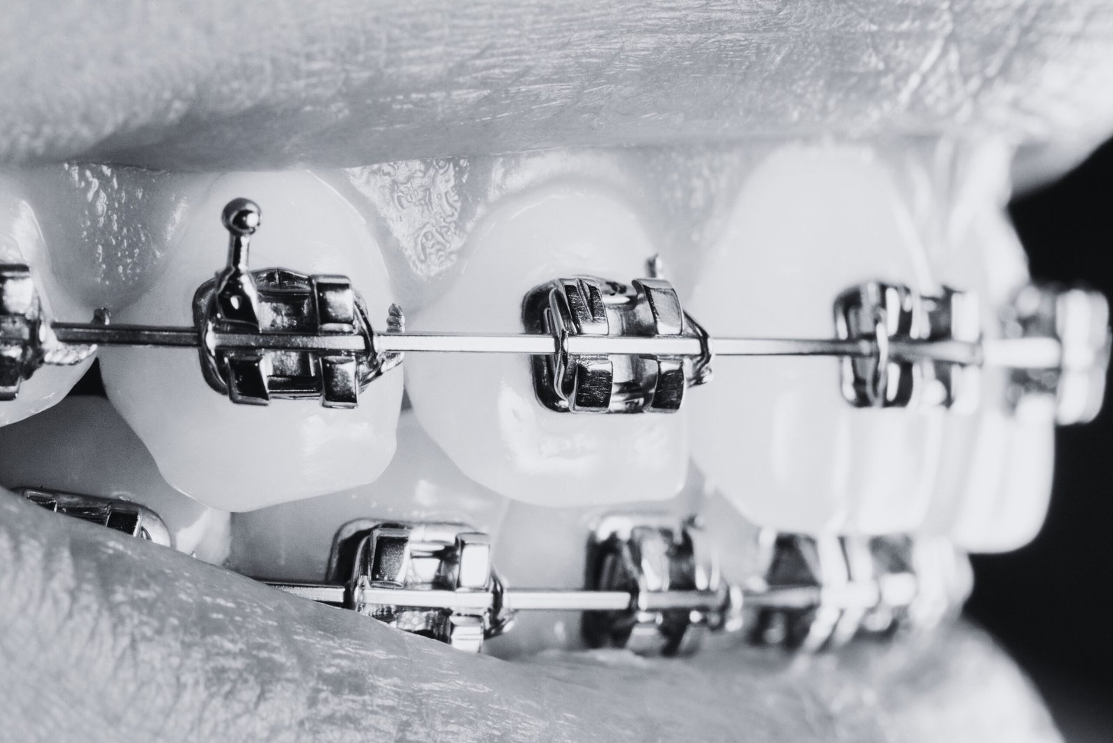
Only orthodontic treatments that meet the specified criteria are accepted. Eligible cases include treatments using fixed appliances, mobile devices, or aligners aimed at correcting occlusion, tooth alignment, and/or arch positioning. If the treatment plan includes orthognathic surgery, the case will be reclassified under Interdisciplinary Oral Rehabilitation. 


Case Description: 

  • A concise clinical case report, not exceeding 3,500 words 
 
  • Include initial orthodontic diagnosis (classification, occlusal and/or aesthetic issues), final diagnosis, treatment plan, type of appliance used, and a brief case summary 
 
  • Presentation must include before/after intraoral and extraoral photos ± radiographic documentation (cephalometric, periapical, or panoramic images) relevant to the treatment 
 
  • Progress photos at key treatment stages are encouraged 


Photo Documentation Guidelines: 

  • Follow standard orthodontic photographic protocol (frontal, lateral, occlusal views) 
 
  • Images must be clear, properly lit, and centered 
 
  • If facial changes are demonstrated (e.g., profile or smile), extraoral before/after photos are recommended 
 
  • All photos must respect confidentiality requirements and exclude any personal identifiers 
 
  • Images must be symmetrical and appropriately cropped 

Only treatments aimed at the prevention, control, and/or regeneration of periodontal conditions are accepted, in accordance with international guidelines (EFP/AAP). Accepted cases may include non-surgical therapy, resective surgery, or regenerative procedures. 


Case Description: 

  • A brief case description, not exceeding 3,500 words 

  • Include initial periodontal diagnosis (staging and grading per EFP/AAP), final diagnosis, treatment plan, therapeutic methods used (including antimicrobials or surgical interventions), and a summary 

  • Presentation must include before/after photos ± relevant radiographs (e.g., periapical, bitewings) 

  • Periodontal charting (probing depths, mobility, recession) before and after treatment is encouraged 


Photo Documentation Guidelines: 

  • Images must clearly depict the condition of soft and hard tissues (e.g., gingiva, attachment, recession, plaque, calculus) 

  • Contrastors and mirror images are recommended to enhance visibility 

  • Photos must be of high clarity, contrast, brightness, and focus 

  • Images should be symmetrical and properly cropped 

  • Follow-up documentation showing clinical improvement is recommended